Methods
Two WBR-based applications were clinically evaluated in Duke University and Vanderbilt University Medical Centers. 39 patients referred for bone SPECT scans were included in this study. Data was acquired following the routine hospital acquisition protocol. Bone SPECT projection data was reconstructed twice: once, using the typical clinical protocol (FBP), and a second time using the relevant WBR reconstruction protocol. All images were blind read. For each reading, a questionnaire was completed, in which the reader was asked to evaluate, on a scale of 1-100, the following parameters:
- Clarity of bone uptake
- Bone to soft tissue ratio
- Overall image quality
- Confidence of interpretation
Results
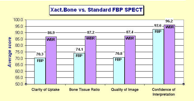
Figure 1. Comparison of the WBR-based Xact.Bone reconstruction with standard FBP reconstruction of SPECT images. The results show a significant advantage of WBR over FBP in all evaluated parameters.
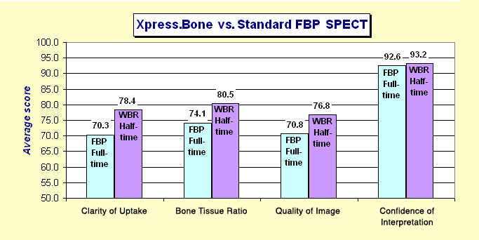
Figure 2. The WBR-based Xpress.Bone reconstruction applied to data collected in half of the standard scan time is compared with standard FBP reconstruction of data collected in full scan time. The Xpress.Bone, utilizing on the average half the counts, scored higher than the routine FBP of the full count data.