Confidence. Much greater visualization for improved diagnosis for higher confidence.
Your patients depend on you. You depend on high quality images. Rest assured that you are giving your patients the best of both worlds. A diagnosis assisted by high quality images, and the lowest possible dose.
Key Benefits
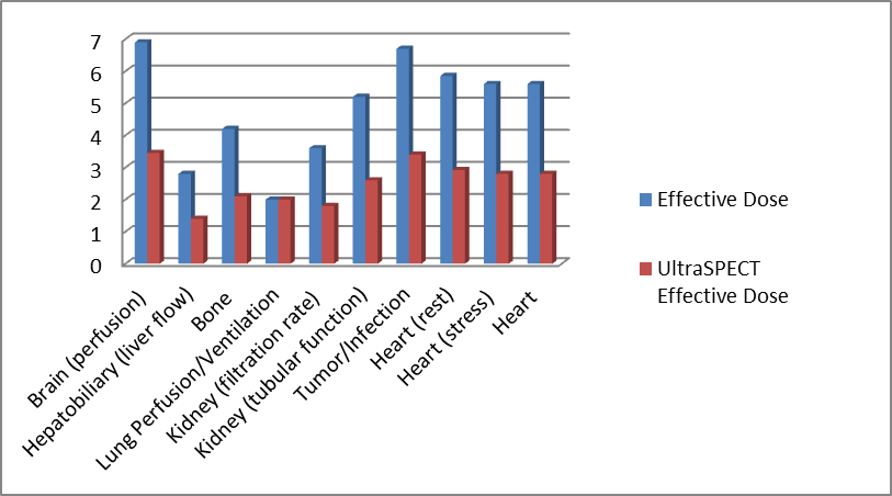
- Image quality: Whether imaging at ½ time or ¼ time or combination of ½ dose and ½ time, your images will be as good, if not better, than your current resolution. UltraSPECT’s exclusive reconstruction algorithms resolve the sensitivity and resolution trade-off problem that is inherent to the gamma camera, and which is critical in SPECT imaging.
- Accurate diagnosis: Accuracy matters. That’s why you can rely on the high image resolution produced by UltraSPECT’s Wide Beam Reconstruction (WBR™ technology. Not only is it equivalent to that of full-time imaging, but the images display higher Signal-to-Noise Ratio with improved background uniformity. And they enable better visualization of endocardial borders and wall motion segments for the most confident diagnosis.
- UltraSPECT technology and products in the literature were clinically validated showing improvement in diagnostic capabilities.
- lesion localization (DePuey, 2008)
http://www.springerlink.com/content/876201n707jg0645/ - visualization of endocardial borders (DePuey, 2008)
http://www.springerlink.com/content/876201n707jg0645/ - diagnostic confidence (Druz, 2011)
http://www.springerlink.com/content/9j45n4h321280510/ - detection of wall motion abnormality (DePuey, 2008)
http://www.springerlink.com/content/876201n707jg0645/
- Standardized protocol (unified protocol): Multi-site, multi-camera organizations benefit from the ability to use the same clinical protocol with consistent results at all sites.
- Adherence to ASNC guidelines: Be a leader and provide added value for your patients by adhering to the ASNC guidelines. Patients are engaged and choosing facilities that will provide them with the lowest dose. Give them a reason to choose you.
Did you know?
As of January 2014, ASNC has announced its low-dose guidelines stating that the cardiology community limit the total radiation exposure to patients undergoing SPECT/PET myocardial perfusion imaging (MPI) tests to an average of less than or equal to 9 mSv (millisieverts) in 50% of studies. Previously, the average was approximately 12 to 13 mSv for stress and rest imaging.
Did you also know?
Study data gathered from a comparison of two facilities has shown that using UltraSPECT software, nuclear medicine imaging departments can achieve the ASNC low dose guidelines while also using their standard imaging protocols.
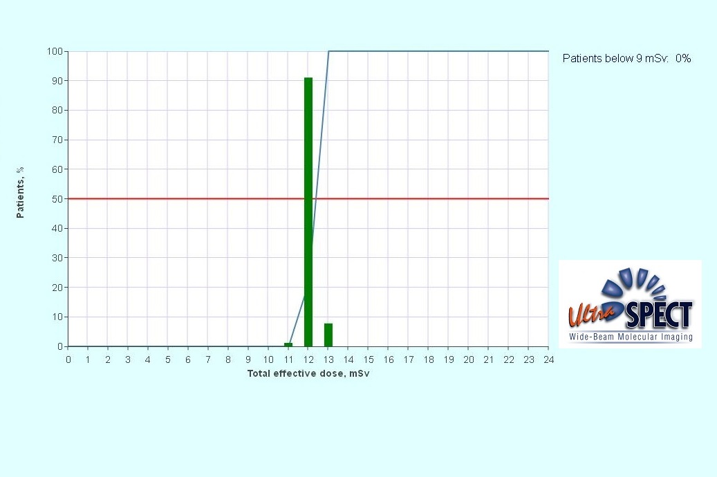
Facility #1: Ffull-dose protocol R/S Mibi with 257 patients. Results: => Below 9mSv – 0%
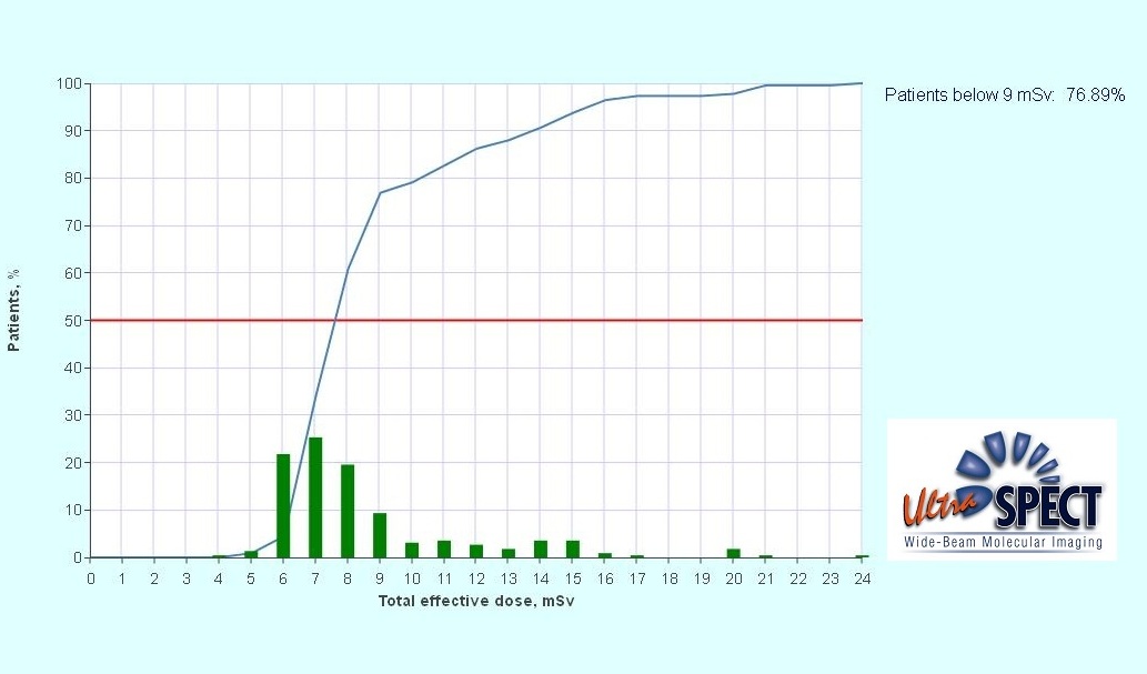
Facility #2: Half-dose protocol with 225 patients (S/R Tl-201 - 2, Two-day high dose - 11, Two-day stress only - 7, Half-dose WBR – 205)
Results: => Below 9mSv – 76.89%
What others are saying:
"Radiation exposure reduction is a global priority and is readily achievable today through a combination of practices that includes innovative software like that from UltraSPECT. MaineHealth is proud to be a leader in meeting the American Society for Nuclear Cardiology dose reduction recommendations as we continue to take steps to implement advanced methods to reduce radiation exposure related to cardiac imaging for the best results for the patients in our care."
Mylan Cohen, MD, Medical Director of Noninvasive Cardiology at Maine Medical Center and former ASNC President..
“What this preliminary data [using UltraSPECT software] shows us is that healthcare facilities do not have to change their standard imaging protocols to meet ASNC guidelines to lower dose. I am looking forward to seeing the great progress that the cardiovascular imaging community can make . . . as proven and viable solutions are applied to lower the dose for the benefit of patients and to continue to advance healthcare.”
E. Gordon DePuey, MD, director of Nuclear Medicine at St. Luke’s-Roosevelt Hospital, New York and past President, ASNC.